

One of the most interesting topics in the field of emotional development research relates to infants´ capacity to express and experience different emotional states. A general question refers to when emotions first emerge in humans life. Approach and withdrawal are two important concepts in the definition of human´s emotions. Both constructs have not typically been viewed as features of infant emotional behavior until late in the first year. However, when looking directly into infants´ brain, researchers have found a different story suggesting that infants experience positive and negative emotions from birth but in a very basic way and not as adults do.
Infants´ emotions are not equivalent to adults emotions
The development of emotions is consistent with infants´ exhibition of many different facial expressions starting during the first year. From birth, infants show an amazing biological capacity to experience basic emotions such as happiness, disgust, and fear. For a long time, researchers have tried to infer infants´ emotional experience just by observing the specific behavioral reactions, such as facial expressions, vocalization, or body movements. Although morphologically stable from 3 months of age (Izard, 1997), infant facial expressions are transient are reflexive mostly reflecting the precocious activity of mirror neurons.
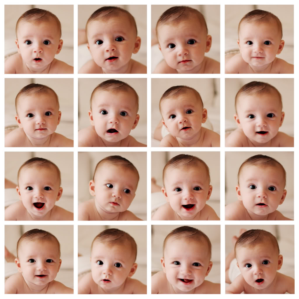
Generally, infant facial expression studies have supported the view that all emotion expressions, especially specific negative emotions, are not psychologically meaningful in the first year of life (Bennett et al., 2002; Sullivan and Lewis, 2003). This has generated much controversy in determining what infants are actually expressing when they smile or tear, in part due to the fact that most of the infants´facial configurations have been derived from adult observations of emotional expressions. What might constitute such facial movement patterns in infants, homologous with the referenced adults´ facial expression is still unclear.
Looking into infants´ brain
To overcome the limitations of behavioral studies, researchers have “looked” into the brain to get more accurate insights. Neuroimaging techniques such as functional magnetic resonance (fMRI) and electroencephalography (EEG) allow researchers to measure infants´neural correlates related to the emotional activity. Consistent evidence indicates that neurons respond to certain patterns of biologically significant information that emanates initially from the mother’s face (Nakamura et al., 2000; Bornstein et al., 2003), and thereby facial expressions have strong biological meaning. Indeed, they serve as key references to extract information about how threatening or safety the environment is for the baby. For instance, happy expressions from the smiling and laughing joyful face of an affective mother indicate no threaten at all.
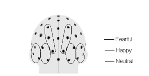
The field of emotional development research has especially focused on the pattern of frontal EEG power (i.e brain energy) in the alpha frequency (Davidson, 2003). Alpha frequency appears early in life and it is considered a brain marker of cortical activation in specific brain regions. A differentiated pattern of alpha EEG frontal asymmetry (FA) has been related to different types of emotions. For example, greater relative left FA activity is associated with approach behaviors such as happiness or anger and with the expression of positive emotions. On the contrary, greater right FA activity is associated with withdrawal behaviors, and with the expression of negative emotions like fear and sadness. Thus, early patterns of FA provide neurobiological information regarding the infant emotional trait dispositions or affective style (i.e. emotionally regulated pattern vs reactivity pattern; Smith and Bell 2010).
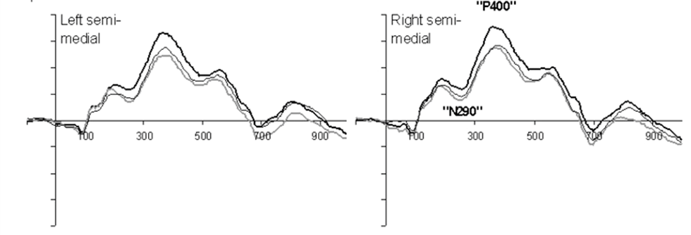
Another common approach to explore brain correlates of infants´emotional experience is by measuring the time course of the neural response after presenting an arousing stimulus (e.g. familiar faces). One physiological measure typically used to investigate whether the brain is processing emotions during perception of faces is the Event-Related Potentials (ERPs) which are defined as patterns of transient changes in neural activity. A large number of studies have identified two important ERPs involved in infants´ human face processing termed by scientists N290 and P400. These markers are elicited when young infants are exposed to different facial expression faces. Interestingly, the electrical brain activity during the presentation facial expressions categories reveals for instance that 7-old-month infants discriminate between happy and fearful expressions but not between angry and fearful expressions (Nelson and De Haan, 1996).
Right brain maturation: what can tell us about the emotional development?
Infants´ emotion can also be tracked in the course of development revealing different brain pathways deployment. Basic emotions such as distress and contentment are displayed early within a maturational period of the right brain, which is dominant in the first three years of human life. The development of an optimal right-brain emotional function greatly depends on early attachment experience between the infant and primary caregiver (Schore, 2001). Either positive or negative, such experiences will have an important influence in the maturation of brain structure, and therefore, the psychological development of the infant. But…how the brain organizes the emergence of emotional processing networks at early ages?
A rudimentary right-hemisphere limbic system starts to be active very early in life, mirroring a reflexive (or tonic) emotional reaction (Ryan et al. 1997). Interestingly, important research has shown that that the growth of the baby’s emotional networks requires a brain to brain interaction which takes place in the context of a positive affective caregiver-infant relationship (Trevarthen, 1993). These dyadic experiences are called ‘affect synchrony’ and operate around two months of age at the earliest, in the context of social interaction (Atzil et al., 2014; Feldman et al. 2007). Within such episodes of coordinated affect, caretakers often engage facial, vocal, and gestural preverbal communications allowing the synchronization of positive affective brain states.

In terms of neural organization, this is expressed as changes in the baseline of infants´ primary brainwaves and EEG rhythms. As suggested by investigators like Tronick and Weinberg (1997), the infant’s emotional system, when coupled with the mother’s, allows for a more adaptative brain organization, thus facilitating the effective development of right-hemisphere socio-emotional circuitries.
Conclusions
Emotions emerge very early in life: first as a natural biological mechanism of adaptation with very basic brain configurations evolving later into more complex and meaningful feelings from a psychological point of view. Without self-consciousness, babies can experience interest, distress, disgust, and happiness from birth, and can communicate these states through facial expressions and body postures as they grow and learn from the environment. At the neural level, infants display a rapid increment of new synaptic connections between frontal lobes and emotional centers showing, during the first year of life, the asymmetrical pattern of frontal activity that characterizes positive and negative emotions in adults. Furthermore, emotional development is influenced by many factors, especially by the baby’s relationships with their primary caregivers.
Reference
Atzil, S., Hendler, T., & Feldman, R. (2014). The brain basis of social synchrony. Social cognitive and affective neuroscience, 9(8), 1193–1202. doi:10.1093/scan/nst105 [Link]
Bennett, D. S., Bendersky, M., & Lewis, M. (2002). Facial Expressivity at 4 Months: A Context by Expression Analysis. Infancy : the official journal of the International Society on Infant Studies, 3(1), 97–113. doi:10.1207/S15327078IN0301_5 [Link]
Davidson RJ. Affective neuroscience and psychophysiology: toward a synthesis. Psychophysiology. 2003;40:655-65 [Link]
Feldman R. Parent-infant synchrony and the construction of shared timing; physiological precursors, developmental outcomes, and risk conditions. Journal of Child Psychology and Psychiatry. 2007a;48(3–4):329–54. [Link]
Halit H, Csibra G, Volein Á, Johnson MH. Face-sensitive cortical processing in early infancy. Journal of Child Psychology and Psychiatry. 2004;45:1228–1234. [Link]
Izard CE. Emotions and facial expressions: A perspective from Differential Emotions Theory. In: Russell JA, Fernandez-Dols, editors. The psychology of facial expression. Cambridge, UK: Cambridge University Press; 1997. pp. 57–77. [Link]
Nelson Charles A., and Michelle de Haan. 1996. Neural Correlates of Infants’ Visual Responsiveness to Facial Expressions of Emotion. Developmental Psychobiology 29, no. 7: 577-595[Link]
Ryan, R.M., Kuhl, J., & Deci, E.L. (1997). Nature and autonomy: An organizational view of social and neurobiological aspects of self-regulation in behavior and development. Development and Psychopathology, 9, 701– 728 [Link]
Schore, A. N. (2001). Effects of a secure attachment relationship on right brain development, affect regulation, and infant mental health. Infant Mental Health Journal, 22(1-2), 7-66 [Link]
Smith, C. L., & Bell, M. A. (2010). Stability in infant frontal asymmetry as a predictor of toddlerhood internalizing and externalizing behaviors. Developmental psychobiology, 52(2), 158–167. doi:10.1002/dev.20427 [Link]
Sullivan MW, Lewis M. Contextual determinants of anger and other negative expressions in young infants. Developmental Psychology. 2003;39(4):693–705. [Link]
Tronick, E. Z., & Weinberg, M. K. (1997). Depressed mothers and infants: Failure to form dyadic states of consciousness. In L. Murray & P. J. Cooper (Eds.), Postpartum depression and child development (pp. 54-81). New York, NY, US: Guilford Press [Link]
Trevarthen, C. (1993). The self-born in intersubjectivity: The psychology of an infant communicating. In U. Neisser (Ed.), The perceived self: ecological and interpersonal sources of self-knowledge (pp.121– 173). New York: Cambridge University Press [Link]
Make a one-time donation
Make a monthly donation
Make a yearly donation
Choose an amount
Or enter a custom amount
I hope you enjoyed reading this post! 🙂
If you’ve found my content helpful, informative, or thought-provoking, I kindly ask that you consider making a donation to help me continue producing new and exciting content for you to enjoy. Your support means the world to me and will allow me to keep bringing you the latest news and scientific insights in the fields of Cognitive Neuroscience, Neurotechnology, Consumer Behavior and Psychology. Any amount, no matter how small, would be greatly appreciated.
Your contribution is appreciated.
Your contribution is appreciated.

1 thought on “Exploring the brain correlates of emotions in babies”